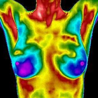
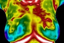
Breast Thermography (or Breast Thermal Imaging) uses a Digital Infrared Camera to detect surface heat; temperature readings are compiled into an image by specialized software for computer analysis, followed by interpretation/evaluation of the images by a trained physician. This screening is best used as a risk assessment and unlike other screenings does not detect calcifications or lumps.
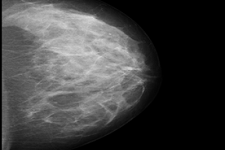
After breasts are compressed between two firm surfaces to spread out the breast tissue, an X-ray captures black-and-white images that are displayed on a computer screen and examined by a radiologist. A Mammogram is the only test that can detect and monitor micro calcifications. Mammography can detect a lump or mass after it reaches 1 centimeter in size. This screening exposes breasts to radiation and is not effective for screening women with dense breasts.
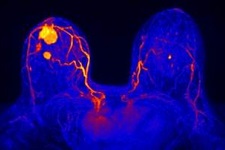
Magnetic Resonance Imaging, also known as an MRI, uses magnets and radio waves instead of x-rays to produce very detailed, cross-sectional pictures of the breasts. For breast MRI to look for a lump or mass, a contrast liquid (called gadolinium) is injected into a vein before or during the scan to show details better. This screening is more expensive than a Mammogram or Ultrasound and is effective for women with dense breasts.
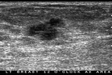
Ultrasound imaging of the breast uses sound waves to produce pictures of the internal structures of the breast. It is primarily used to help diagnose breast lumps or other abnormalities your doctor may have found during a physical exam, mammogram or breast MRI. The primary use of breast ultrasound is to help diagnose breast abnormalities detected by a physician during a physical exam (such as a lump) and to characterize potential abnormalities seen on mammography or breast magnetic resonance imaging (MRI).
Ultrasound imaging can help to determine if an abnormality is solid (which may be a non-cancerous lump of tissue or a cancerous tumor), fluid-filled (such as a benign cyst) or both cystic and solid. Ultrasound is safe, noninvasive and does not use radiation.
Basically an extension of a digital mammogram, the breast is compressed once and a machine takes many low-dose x-rays as it moves over the breast. The images are then combined into a 3-dimensional picture. This screening exposes breasts to more radiation than a mammogram.